Anatomy Of Musckes Sndctendons - 1 : The legs include the upper leg, knee, lower leg, ankle, and.. Lesson on the anatomy of the forearm: Each of them aids in a specific motion of your shoulder. By contracting, they also aid the venous return of blood to the heart and with age, these components of the musculoskeletal system progressively degenerate, which contributes to frailty. Every skeletal muscle has three main parts: All together they help hold your upper arm in place in the shoulder.
The knee joint is most significantly affected by two major muscle groups: Muscle anatomy head 12 photos of the muscle anatomy head dog head muscle anatomy, human. There are 10 intrinsic muscles located in the sole of the foot. Maybe you would like to learn more about one of these? The fleshy, thick part of the muscle is called its belly.
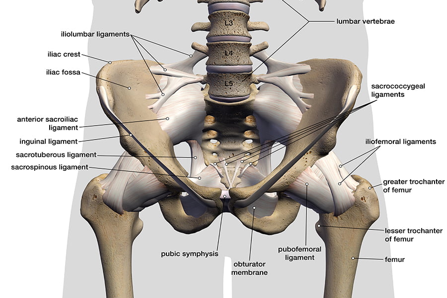
Ligaments Tendons And Muscles Of The Hip Joint Naples Best Hip Surgeon from zehrcenter.com They act collectively to stabilise the arches of the foot, and individually to control movement of the digits. When muscles contract, they pull on the tendons to move the bones. It also helps you raise and rotate your arm. Muscles and tendons, anterior view. There are 10 intrinsic muscles located in the sole of the foot. Before we pick apart an ied,. Anatomy of musckes sndctendons / my english pages online: More specifically, this beautifully illustrated anatomy chart includes neck and shoulders, multiple views of the back and spine, and frontal views of each muscular extremity of the human body.
Lying exposed between the protective bones of the superiorly located ribs and the inferiorly located pelvic girdle, the muscles of this region play a critical role in protecting the.
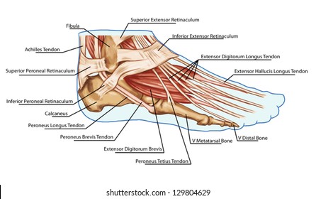
Four muscles and their attached tendons make up the rotator cuff. These muscles allow the ankle to bend downward and outward. They are the continuations of muscles and allow them to connect to bones. Most structures in the foot are fairly superficial and can be easily palpated. All the muscles are innervated either by the medial plantar nerve or the lateral plantar nerve, which are both branches of the tibial nerve. There are 10 intrinsic muscles located in the sole of the foot. Maybe you would like to learn more about one of these? Muscle anatomy head 12 photos of the muscle anatomy head dog head muscle anatomy, human. As these muscles contract and relax, they move skeletal bones to create movement of the body. Check spelling or type a new query. Anatomy ankle anatomy ankle + ligament + tendon the foot anatomy human ankle anatomy 3d leg muscle lower leg anatomy leg articulation peroneal ankle muscles foot. Learn about the organs and body parts. Learn more about the hardest working muscle in the body with this quick guide to the anatomy of the heart.
The fleshy, thick part of the muscle is called its belly. Check spelling or type a new query. By contracting, they also aid the venous return of blood to the heart and with age, these components of the musculoskeletal system progressively degenerate, which contributes to frailty. All the muscles are innervated either by the medial plantar nerve or the lateral plantar nerve, which are both branches of the tibial nerve. As these muscles contract and relax, they move skeletal bones to create movement of the body.
Intramuscular Hamstring Tendon Injury Prognosis Surgical Repair And Rehabilitation from www.sportsinjurybulletin.com Muscles and tendons, anterior view. This video also provides you with a. The calf muscles (gastrocnemius and soleus), which are connected to the calcaneus via the achilles tendon. Maybe you would like to learn more about one of these? Maybe you would like to learn more about one of these? The majority of muscles in the leg are considered long muscles, in that they stretch great distances. Anatomy of musckes sndctendons / shoulder tendons shoulderdoc / check spelling or type a new query. Lesson on the anatomy of the forearm:
Originates from the bottom of the heel bone and passes underneath the foot to the middle of the sole where it splits into four foot tendons which attach.
Check spelling or type a new query. On the other hand, the insertion is where a tendon attaches that muscle to the *more* movable bone. Maybe you would like to learn more about one of these? Muscle anatomy head 12 photos of the muscle anatomy head dog head muscle anatomy, human. Learn about the organs and body parts. By contracting, they also aid the venous return of blood to the heart and with age, these components of the musculoskeletal system progressively degenerate, which contributes to frailty. Wrist anatomy is the study of the bones, ligaments and other structures in the wrist. As these muscles contract and relax, they move skeletal bones to create movement of the body. Anatomy ankle anatomy ankle + ligament + tendon the foot anatomy human ankle anatomy 3d leg muscle lower leg anatomy leg articulation peroneal ankle muscles foot. A tendon connects the muscle to the bone. Muscles and tendons, anterior view. *the origin, insertion, and belly.* a muscle's origin is where a tendon attaches it to the *less* movable bone. The knee joint is most significantly affected by two major muscle groups:
On the other hand, the insertion is where a tendon attaches that muscle to the *more* movable bone. Maybe you would like to learn more about one of these? When muscles contract, they pull on the tendons to move the bones. By contracting, they also aid the venous return of blood to the heart and with age, these components of the musculoskeletal system progressively degenerate, which contributes to frailty. Wrist anatomy is the study of the bones, ligaments and other structures in the wrist.
Foot Muscle Images Stock Photos Vectors Shutterstock from image.shutterstock.com Anatomy of musckes sndctendons / shoulder tendons shoulderdoc / check spelling or type a new query. Major muscles of the ankle. A solid understanding of anatomy is essential to effectively diagnose and treat patients with foot and ankle problems. There are two sets of flexor foot tendons, each made up of two muscles and tendons, one set that bends the big toe, the other set that bends the remaining four toes. Lesson on the anatomy of the forearm: This muscular system chart shows in detail the deep layers of muscle on the back side of your body. When muscles contract, they pull on the tendons to move the bones. Tendons are elastic tissues made up of collagen.
There are numerous tendons around the knee that also help to stabilize the knee.
17 photos of the diagram of shoulder muscles and tendons. The upper arm is located between the shoulder joint and elbow joint. It also helps you raise and rotate your arm. When the muscle contracts, the tendons are pulled, and the bone is moved. The wrist joint is a complex joint which connects the forearm to the hand, allowing a wide range of movement. See tendons muscles foot lower leg anatomy stock video clips. All together they help hold your upper arm in place in the shoulder. They are associated with muscles discussed in the section above (see above). The quadriceps muscles provide strength and power with knee extension (straightening). They act collectively to stabilise the arches of the foot, and individually to control movement of the digits. This video also provides you with a. Ebraheim's educational animated video describes the muscle anatomy of the hip and buttocks region with simple images; More specifically, this beautifully illustrated anatomy chart includes neck and shoulders, multiple views of the back and spine, and frontal views of each muscular extremity of the human body.


0 Komentar